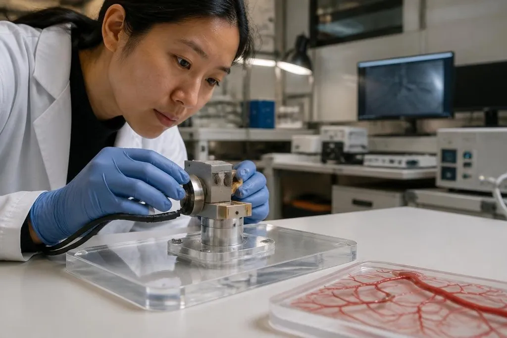
Medical micro-robots could move through blood vessels, release drugs, or reach tissue that tools struggle to access. Their small size creates the same problem that makes them useful: there is little room for power, sensors, control hardware, or a way to recover the robot.
- Steering usually depends on equipment outside the body.
- Medical imaging must show the robot and the tissue around it.
- Safe use requires a clear plan for retrieval, breakdown, or removal.
Steering inside the body
A doctor needs to know where the robot is and control its motion after it enters the body. Many designs use external magnetic fields, ultrasound, or light to move the robot because a battery and radio link may not fit inside its body.
That approach shifts the problem to the equipment around the patient. Magnetic steering needs a field that reaches the target with enough force. Ultrasound can push or heat material, while light works only where it can reach the robot.
Blood flow, breathing, heartbeat, and soft tissue can move the robot away from its planned route. The robot also needs to stop at the right place. A small error may leave a drug dose beside the target instead of inside it, or send the device into a smaller vessel where it cannot turn.
Power and movement
A micro-robot has little space for an energy source. Designs that move by magnetic force can avoid carrying a motor, but the patient then remains connected to a large control system. That limits movement to places where the system can create a useful field.
Some robots swim with tiny screws, flexible tails, or changes in shape. Each method faces resistance from the fluid or tissue around it. A design that moves well in a lab dish may slow down in blood, mucus, or a narrow passage filled with bends.
Battery-free movement also leaves open questions about control during a pause. If the robot loses contact with its magnetic or sound field, it may drift until the operator finds it again.
Seeing the robot
A clinician cannot safely guide a device they cannot locate. Medical imaging can help, but each method shows a different part of the problem. X-ray can track dense materials, ultrasound can show motion in soft tissue, and magnetic resonance imaging can show anatomy while also affecting magnetic devices.
Visibility against the body matters. That calls for markers or materials that appear clearly without blocking the view of the target. Imaging can also add time, radiation, heat, or equipment limits, depending on the method and procedure.
A clear image in a lab does not prove that a medical micro-robot can be tracked during a procedure. Reporting linked through Robot 24 can place the device, imaging method, test setting, and result beside the claim before clinical testing begins. The next check is whether the robot can leave the body safely.
Safety after the task
A medical device needs a known fate after it reaches its target. The robot may need to leave the body intact, dissolve into approved materials, or remain in place for a set period before removal. Each choice adds tests for toxicity, infection risk, blockage, and tissue damage.
A stuck robot creates a separate problem. The care team needs a recovery method that works if the device stops moving, loses its coating, or reaches the wrong vessel. The plan must also account for fragments if the robot breaks apart.
Sterilization can change the robot's shape, coating, or magnetic behavior. Repeated production adds another test: every unit must behave close enough to the one used in the safety study. Small changes matter when a device works inside a vessel narrower than a fingertip.
What must happen before wider use
The strongest lab result does not answer every medical question. A useful test plan should connect the robot's design to a specific procedure, target, imaging method, and recovery path.
Use this checklist when judging a medical micro-robot project:
- Target site: Name the tissue or vessel and state how the robot reaches it.
- Control method: Identify the field, sound, light, or other force that moves it.
- Tracking method: Show how the clinician sees position, speed, and orientation.
- Failure response: Describe what happens after drift, power loss, blockage, or breakage.
- Material safety: Give the coating, body material, sterilization method, and removal plan.
- Clinical proof: Separate work in a dish or animal model from results in people.
A project that answers these points has a clearer path to testing. A project that shows only motion leaves the hardest part unanswered.
I'd wait for evidence from realistic anatomy and a tested recovery plan before treating any medical micro-robot as ready for routine care. The field's next useful milestone is a repeatable procedure in which doctors can see, steer, stop, and remove the device when the plan changes.